Virtual Prostate Visualization & Computer-Aided Detection
Method, system, and computer-readable medium for detecting a disease of a prostate
Prostate cancer (CaP) is the most commonly diagnosed cancer among males in Europe, and is the second leading cause of cancer related mortality for this same group. Although it is such a common cancer, diagnosis methods remain primitive and inexact. Detection relies primarily on the use of a simple blood test to check the level of prostate specific antigen (PSA) and on the digital rectal examination (DRE). If an elevated PSA level is found, or if a physical abnormality is felt by the physician during a DRE, then biopsies will be performed. Though guided by transrectal ultrasound (TRUS), these biopsies are inexact, and large numbers are often necessary to try and retrieve a sample from a cancerous area.
This technology involves methods and processes for non-invasive examination of the prostate (and other glands) using interactive visualization and computer-aided detection (CAD) of cancer, called the Virtual Prostate, leading to a better visualization, diagnosis and therapeutics of prostate cancer, and consequently reducing mortality from prostate cancer. The technology employs scanning (typically using MRI of the pelvis or endorectal) and computer processing and visualization.
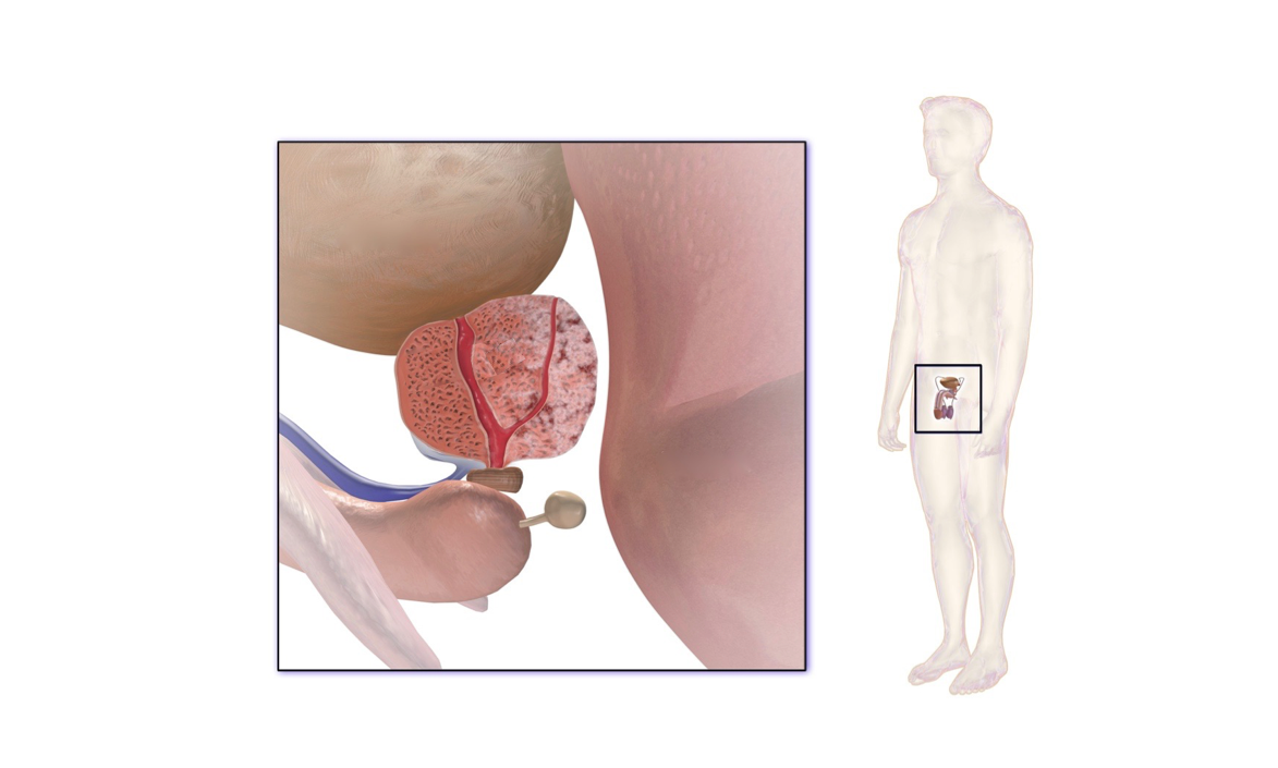
 Please note, header image is purely illustrative. Source: BruceBlaus, Wikimedia Commons, CC BY-SA 4.0, edited.
Prostate cancer is the number two cancer killer among males. This technology provides new methods of imaging the prostate and utilizing this information as needed, leading to a better visualization, diagnosis and therapeutics of prostate cancer in a non-invasive manner. As there are no existing methods to localize prostate cancer, this technology is a breakthrough and a pioneering technology.
- Receiving an image dataset acquired with at least one acquisition mode - Segmenting a region of interest including the prostate from the dataset - Applying conformal mapping to map the region of interest to a canonical shape - Generating a 3D visualization of the prostate using the canonically mapped dataset - Applying computer aided detection (CAD) to the canonically mapped volume to detect a region of disease of the organ
Patented
8995736
Available for license.
Development partner,Commercial partner,Licensing
Patent Information:
| App Type |
Country |
Serial No. |
Patent No. |
Patent Status |
File Date |
Issued Date |
Expire Date |
|