Wide-Field, Coherent Scatter Imaging for Improved Mammography Screening
A method for enhancing conventional radiographic imaging systems by capturing and analyzing otherwise discarded information contained in coherent scatter patterns.
Mammography is a very reliable tool for reducing mortality rates due to breast cancer through early detection. However, the complexity of structures in the breast make it difficult for health care professionals to distinguish between cancerous and otherwise healthy tissues using current medical imaging techniques. This necessitates the use of invasive procedures, such as biopsies, to definitively diagnose the patient. Approximately 80% of biopsies performed on the basis of a mammogram screening are benign, indicating that the current system causes unnecessary patient trauma and wastes critical healthcare system resources. In addition, conventional screenings may fail to detect micro-calcifications and other signs of cancer, which could lead to critical delays in treatment.
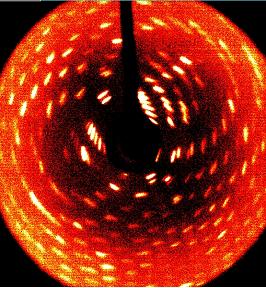
This patented technology provides a method to detect the coherently scattered radiation simultaneously with the normal image by adding additional absorption grids and detectors to existing slot scan imaging systems. These grids are directed at an angle to the main beam, so they transmit the scattered radiation from one particular angle, and thus only from particular tissue types. Furthermore, the intensities collected through the coherent scatter pattern can be projected in three dimensions and allow for localization of specific tissues on the conventional image. This technique can be applied to any radiographic energy for discriminating between tissue types and has the potential to provide much higher degrees of accuracy in the diagnosis of disease.
The benefit of using coherent scatter imaging in addition to conventional methods arises from the fact that the nanoscale structure of tissues and inorganic materials can be revealed by distinguishing and mapping the diffracted x-rays, which occur at predictable angles for specific materials. With this understanding, new systems can be developed, or modifications can be made to conventional slot scanning systems already available at most clinical medical offices.

- Increased specificity and sensitivity of radiological imaging techniques, leading to earlier and more reliable detection.
- Decreased false positive readings and a decrease in unnecessary testing and trauma for patients.
- No additional patient exposure to radiation required to collect higher quality images.
- Mammography screening and improved detection of breast cancer.
- Medical applications in veterinary services or animal studies.
This invention has been granted U.S. Patent Number 7,646,850.
University at Albany’s Office of Innovation Development and Commercialization is currently seeking academic and corporate partners who may be interested in participating in development, licensing, or other commercialization of this technology.
The development of this innovation was funded in part by the US Army Medical Research Materiel Command under grant number DAMD17-02-1-0517.
Patent Information:
| App Type |
Country |
Serial No. |
Patent No. |
Patent Status |
File Date |
Issued Date |
Expire Date |
|