Accurate Computing Tissue Percentages Inside Each Medical Image Voxel
Computation of percentages of different tissue types inside a medical image voxel tissue for medical diagnosis/detection methods.
Image segmentation play an important role in quantitative analysis of medical images for various clinical applications. Traditional image segmentation algorithms cluster all image voxels into several groups and assign a unique label to each group. However, not all voxels in each labeled group contain the same tissue type.
The algorithm developed by Stony Brook University researchers allows for the computation of the mathematically and statistically exact percentages of different tissue types inside a medical image voxel. From the tissue percentages, the tissue properties for medical diagnosis and development of computer aided detection/diagnosis methods can be found. This technology describes a method and apparatus developed for both automated and interactive operations on an image model of the human body. The tissues of the body are segmented and displayed by colors and semi transparency modes. The operations on the tissues are interactive in real time.
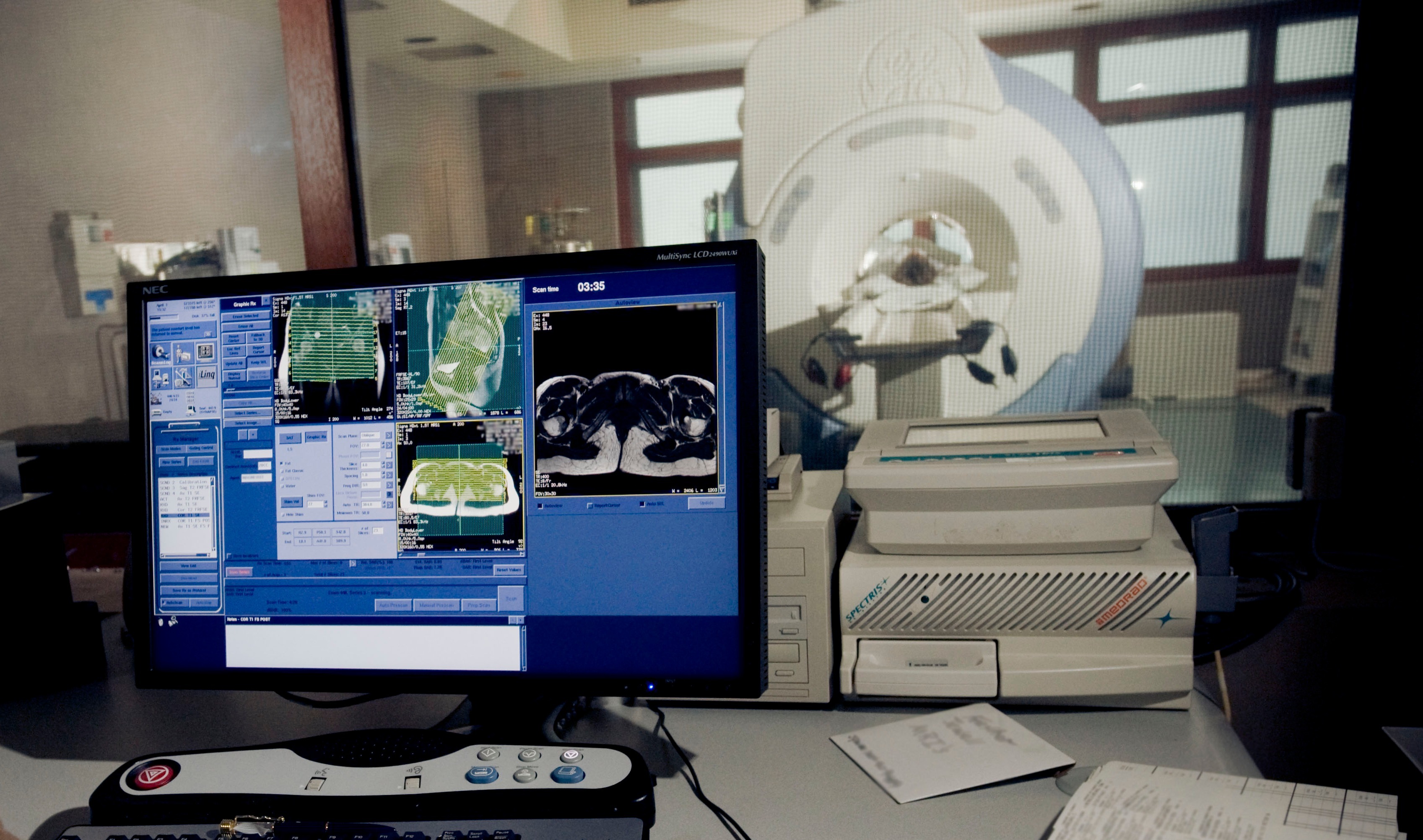 Please note, header image is purely illustrative. Source: U.S. Air Force photo by Master Sgt. Keith Brown, public domain.
This is the first coded software on currently available, inexpensive computer systems to do both automated and interactive surgical planning for congenital aural atresia. It is clinically tested by a surgeon.
- Digital Medical Imaging Software - Magnetic Resonance Imaging
This technology is available for sponsored research.
Seeking investment
Patent Information:
| App Type |
Country |
Serial No. |
Patent No. |
Patent Status |
File Date |
Issued Date |
Expire Date |
|