Noninvasive Epithelial Cancer Diagnosis and Grading
A novel OCT system to acquire high resolution, in-depth optical images of living tissue samples.
Optical Coherence Tomography (OCT) works by projecting a laser into tissue. When the light is reflected back, the OCT microscope uses an optical interferometer to detect a proportion of the light that is least back scattered. The system discards the multiple scattered light and uses depth-resolved and intensity information to generate an image. Unfortunately, most biological tissue, such as urinary bladder tissue, is optically opaque. Not only does it absorb the light but scatters it in the optical and near-infrared wavelength range, which makes it almost impossible for existing OCT systems to acquire high resolution, in-depth optical images.
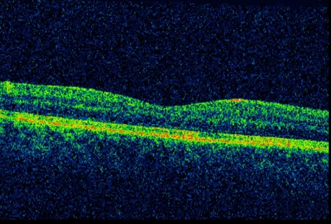
Researchers at Stony Brook University have developed an OCT system that can acquire high resolution, in-depth optical images of a living tissue sample with a specifically chosen time-lapse frame averaging approach. His breakthrough is in the way the ultrahigh-resolution OCT technique takes advantage of micromotion in living biological tissue to effectively reduce speckle noise. This simultaneously reveals the sub-cellular details and imaging of the underlying tissue morphology ? connective tissue and muscle in bladder ? at up to 700 micrometers of depth without focal tracking. This OCT system is ideally suitable for endoscopic imaging applications, such as cancer diagnosis and grading. For more details: In vivo bladder imaging with microelectromechanical-systems-based endoscopic spectral domain optical coherence tomography. Wang Z. et al. J Biomed Opt. 2007 May-Jun;12(3):034009
 Please note, header image is purely illustrative. Source: uguronder, Flickr, CC BY 2.0
- Live sub-surface images at subcellular resolutions (3 micrometers laterally, 1.8 micrometers axially). - No preparation of the sample required. - No sample damage. - Integrates with micro-electro-mechanical system (MEMS)-based laser scanning endoscope for subcellular imaging diagnosis.
- Non-invasive diagnosis and grading of epithelial cancers - Optical biopsy - Image-guided, minimally-invasive surgery
Patent: 8,948,846
Avaialble for Licensing
Patent Information:
| App Type |
Country |
Serial No. |
Patent No. |
Patent Status |
File Date |
Issued Date |
Expire Date |
|