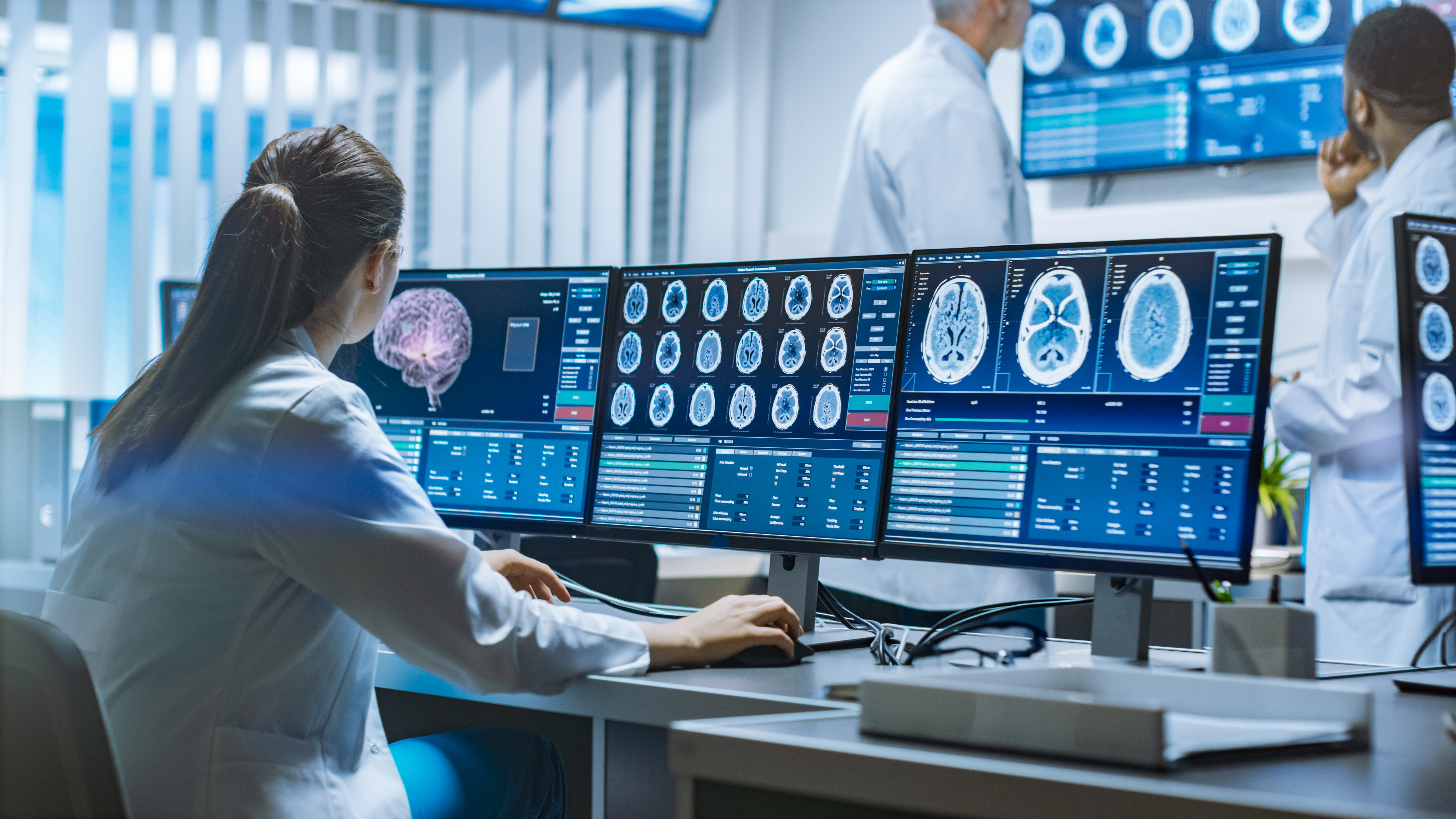
Enables high-quality emission computers tomography (ECT) whild reducing the patient's radiation exposure.
Emission computed tomography (ECT) is an important scientific and medical imaging technique. Clinical applications of ECT include detecting, staging and monitoring response to cancer therapy; detection and risk stratification of cardiovascular diseases; mapping regional blood flow in the brain; bone scans; pulmonary ventilation/perfusion scans; renal scans and many other applications. There is a great need to reduce radiation dose to the patients undergoing ECT examinations. However, dose reduction implies using less ionizing radiation, i.e. fewer gamma photons, which in turn leads to increased noise in the data and in the images reconstructed with conventional methods. Clearly, good quality ECT reconstructions from low-dose ECT examinations, ergo high-noise data, are in high demand. This demand may be met by applying novel advanced ECT reconstruction methods that subdue the noise while preserving diagnostic image quality.
This technology uses a computer to reconstruct transaxial images of the patient from projection data. The reconstruction process estimates mean radiotracer activity distribution inside the body of a patient. It is governed by molecular and/or functional processes and allows clinicians to arrive at diagnoses. The algorithm reconstructs the expected activity distribution, while suppressing noise and preserving the spatial resolution in the estimation. This method demonstrates superior performance vs. standard-of-care when the observed data are very noisy or incomplete. It allows clinicians to perform the clinically acceptable ECT examinations with a radiation dose that is two to six times lower than presently used for the standard-of-care ECT imaging.

• Allows lower radiation dose for patients undergoing ECT examinations without compromising the obtained images quality.
• Provides good quality tomographic image reconstructions under high noise conditions.
Emission computed tomography (ECT). This is widely used for many medical applications, including:
• Detecting, staging and monitoring response to cancer therapy.
• Detection and risk stratification of cardiovascular diseases.
• Mapping of regional blood flow in the brain.
• Bone scans.
• Pulmonary ventilation/perfusion scans.
• Renal scans
… and many others.
This technology is protected by
U.S. Patent 9,460,494, “Methods and systems for inverse problem reconstruction and application to ECT reconstruction”
TRL 4 – Technology validated in lab
This technology is available for licensing.
This technology would be of interest to any organization involved in ECT, including:
• Medical imaging equipment manufacturers
• Hospitals and medical laboratories
• Educational and research laboratories