Radiation Therapy with Orthovoltage X-rays and a Special Collimator
Method of therapeutic radiation delivery for a specific target using a multi-aperture collimator within a trajectory of orthovoltage x-rays
Current radiation therapy is carried out using mostly high energy x-rays. The current MeV x-rays being used have good qualities such as high tissue penetration and a skin-sparing effect. Despite this, several drawbacks are associated with this method; most importantly, the tissue surrounding the target receives radiation, which is damaging to the tissue. These high energy x-rays interact in a scattering mode rather than a photoelectric one, which would be more confined within the target. To address this issue, a grid theory was developed using orthovoltage x-rays. These techniques would be beneficial if they offered tissue-sparing to healthy tissue near the target tissue; without this quality, it doesn't solve the issue of damaging normal tissue. A need for a method of radiotherapy using orthovoltage x-rays for effectively treating tumors is necessary, but the skin and tissue surrounding the target tissue must be spared in order for this method to be effective.
This technology is a system and method for treating a tumor while sparing the surrounding healthy tissue; it uses orthovoltage x-ray radiation. This technology also discloses how to control the tissue depth reached by the radiation, which is essential in order to protect surrounding healthy tissue. This system uses a multi-aperture collimator near the surface of the skin; this lies within a trajectory of radiation, and generates an array of minibeams, which are designed to merge, forming an effective radiation beam.
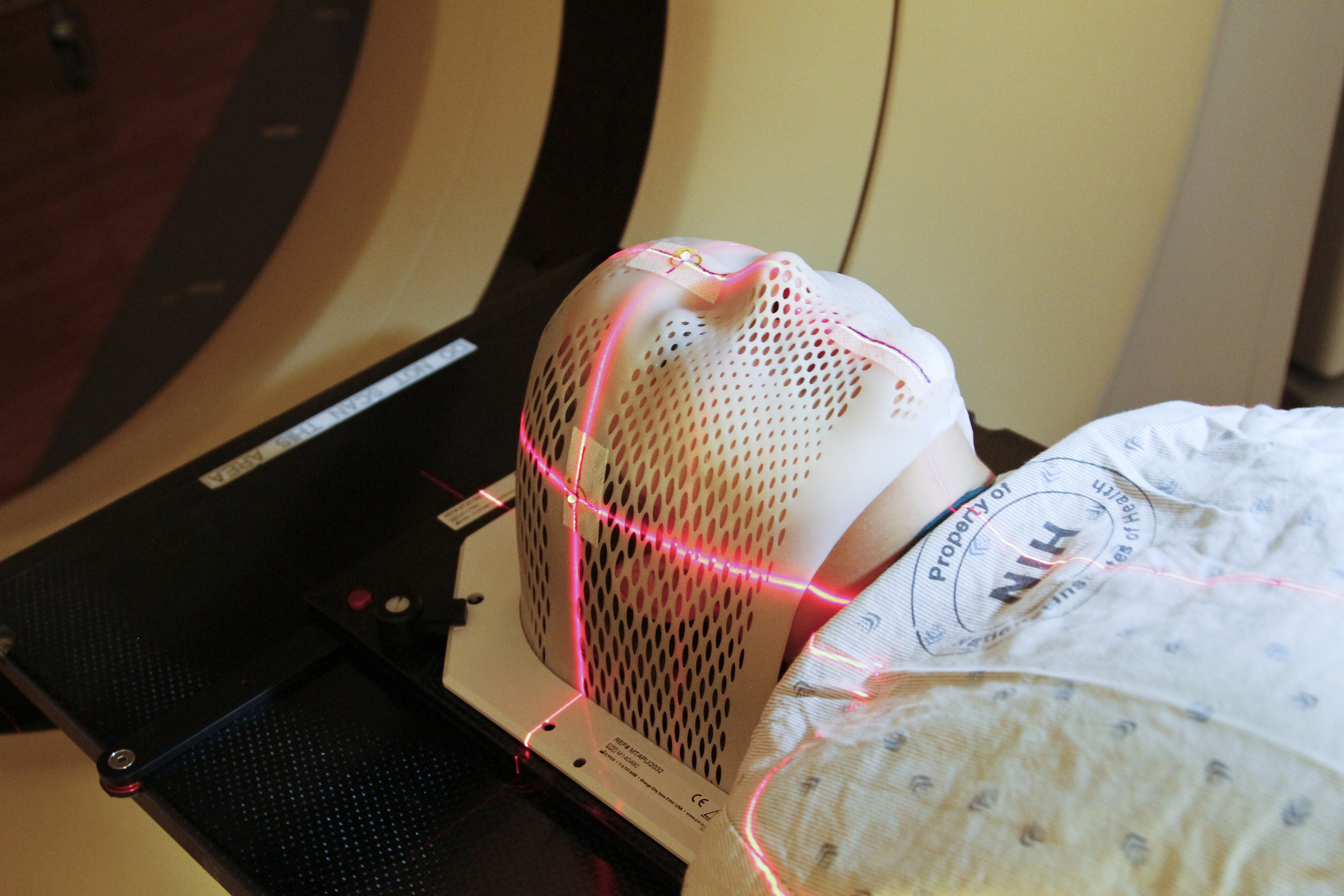
 Please note, header image is purely illustrative. Source: Daniel Sone, NCI, public domain.
- Low-cost radiotherapy - Compact solution for radiotherapy- Controlled beams of radiation to minimize damage of surrounding tissue - Tissue depth can be varied from around 1 cm to 10 cm - Beam broaden to less than 1.0 mm during merging process - Angular position of the x-rays can be changed to hit target from different angles
- Treatment of tumors - Treatment of neurological targets, such as: - Brain tumors - Disorders, such as epilepsy and alzheimers
Patented
10124194
Available for partnership.
Development partner,Commercial partner,Seeking investment
Patent Information:
| App Type |
Country |
Serial No. |
Patent No. |
Patent Status |
File Date |
Issued Date |
Expire Date |
|