Labeling Microtubes in Live Cells
A technique that allows live visualization of microtubules without genetic manipulation or perturbing protein function
Microtubules are a fundamental component of the cytoskeleton of eukaryotic cells, and the observation of microtubule dynamics in live cells using fluorescence microscopy is of critical importance in studying cytoskeleton biology. Microtubules are part of the mitotic spindle in mammalian cells, and their proper organization is essential for normal cell processes. Although microtubules perform heterogeneous tasks in cells, their basic structure is uniform. The core of the microtubule is entirely composed of tubulin, a 100 kDa heterodimer, that assembles to form dynamic cylin-drical structures. Current methods of observation rely on expression of tubulin fused with a fluorescent protein, introducing exogenously labeled tubulin, or using fluores-cently labeled microtubule binders such as taxol.
The present technology provides a technique that can be employed to visualize microtubules in live cells without genetic manipulation or perturbing the protein function. Specifically, this technique exploits the mechanism of tyrosination/detyrosination cycle, a posttranslational modification specific to the C-terminus of α- tubulin, to covalently attach a chemical probe to it. Cells are first grown in medium supplemented with a bioorthogonal amino acid, such as 3-formyltyrosine (3fY). 3-Acetyl tyrosine or tyrosine hydrazide. These un-natural amino acids readily enter the cell and can be appended to the C-terminus of α-tubulin by endogenous tubulin tyro-sine ligase, but are not generally incorporated into proteins produced by the normal protein synthesis mechanisms, thus providing specificity based on the post-translational modification enzymes. Cells are then treated with a suitably derivatized fluorophore and reacts specifically with a reactive chemical site on the bioorthoginal amino acid.
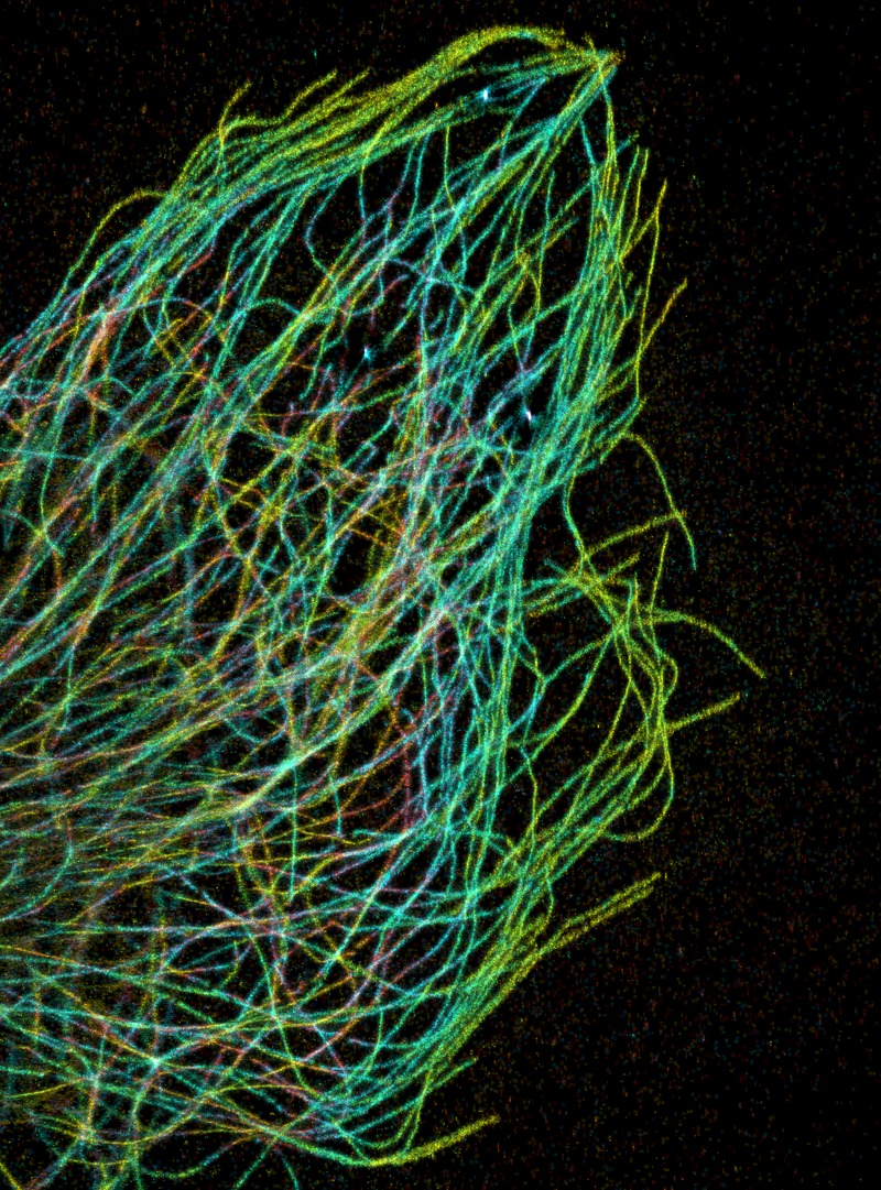 https://en.wikipedia.org/wiki/Microtubule#/media/File:Microtubules_in_the_leading_edge_of_a_cell.tif
- Permits study of microtubule dynamics—formulation of potential chemotherapeutics & assistance in determining mechanism of microtubule drug action.
- No genetic manipulation involved.
- Site-specific labeling.
- Cell morphology, microtubule network and cell viability re-mains unaltered (24 hours).
- Variety of commercially available probes can be used for labeling
US 10,071,990
Patent Information:
| App Type |
Country |
Serial No. |
Patent No. |
Patent Status |
File Date |
Issued Date |
Expire Date |
|